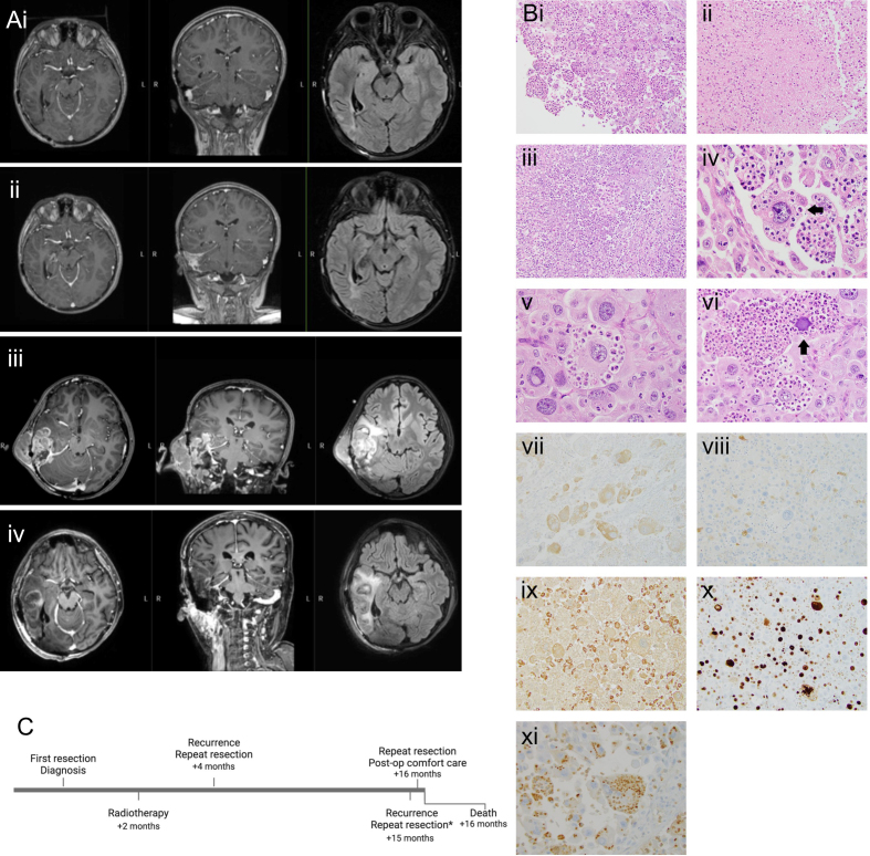
Figure 2.
(A) MRI of a heterogeneously enhancing mass with perilesional edema and progression over time including T1 post-contrast and T2 flair. (i) 12 months post-diagnosis. (ii) 14 months post-diagnosis. (iii) 15 months post-diagnosis, pre-operatively. (iv) 15 months post-diagnosis, post-operatively. (B) Histology. (i) Epithelioid tumor cells sometimes have a “ball of neutrophils” appearance - 200X. (ii) Bland necrosis - 200X. (iii) Necrosis with abundant neutrophils - 200X. (iv) Mostly viable neutrophils within intracytoplasmic vacuolar spaces of cell (arrow) - 600X. (v) Varying degrees of engulfed neutrophil degeneration - 600X. (vi) Degenerate basophilic nucleus in tumor cell (arrow) - 400X. (vii) BRAFV600E immunopositive tumor cells - 200X. (viii) S100 immunonegative tumor cells; S100 immunopositive small histiocytes - 200X. (ix) CD68 immunonegative tumor cells; CD68 immunopositive small histiocytes - 200X. (x) Ki-67 immunopositivity in many tumor nuclei - 200X. (xi) Myeloperoxidase immunostaining highlights the predominantly intracytoplasmic localization of neutrophils - 400X. (C) Timeline of the patient’s clinical course. Histology in (B) is from the patient’s resection at 15 months post-diagnosis.

