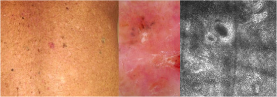
FIGURE 2.

(A, B, C): Clinical, dermoscopic, and confocal presentation of basosquamous carcinoma of the back. At clinical examination, an erythematous macule located on the back of an 87‐year‐old patient (A). Dermoscopy revealed the presence of whitish structureless areas, superficial scales with blood spots in the keratin mass together with blue‐grey blotches in the upper part of the tumor on a background characterized by a polymorphous vascular pattern (B). RCM analysis showed an atypical honeycomb pattern, an inflammatory infiltrate with bright tumor islands and cleft‐like dark spaces (C).
