
Figure 4.
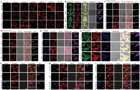
CyR‐SS‐L PSs mainly localize in mitochondria in cancer cells. A) MCF cells were treated with 1 × 10⁻⁶ m of CyH‐SS‐L, CyBr‐SS‐L, or CyI‐SS‐L and the fluorescence signals generated by the cells at different processing times were collected by CLSM. B–E) The subcellular localization of the PPSs in KB‐C2, KB‐3‐1, MCF7, and MCF7/taxol cells incubated with 1 × 10⁻⁶ m CyI‐SS‐L for 2 h was detected using CLSM. F) Detection of the mitochondria localization of CyBr‐SS‐L, CyI‐SS‐L and CyH‐SS‐L in 4T1 cells. G–I) Detection of Golgi apparatus, lysosome, and nuclear localization of CyH‐SS‐L, CyBr‐SS‐L, and CyI‐SS‐L, respectively in 4T1 cells. For MitoTracker green, LysoTracker green, and GolgiTracker green: (λ ex = 488 nm, λ em = 500–580 nm), for nucleus: (λ ex = 350 nm, λ em = 400–500 nm), for CyR‐SS‐L (λ ex = 640 nm, λ em = 650–730 nm). Scale bar = 50 µm.