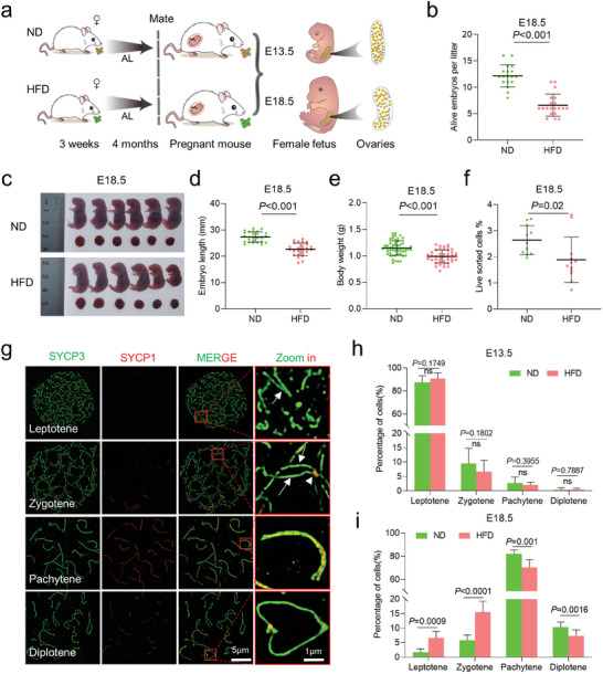
Figure 1.

Maternal obesity impairs fetal development. a) Diagram illustrating the high‐fat diet (HFD) model in ICR female mice. Normal diet (ND) mice were provided with a standard diet and HFD with high‐fat diet. Mice were subjected to either ND or HFD feeding for a duration of 4 months, starting at 3 weeks of age. Subsequently, they were mated with ND males, and the fetuses were harvested at embryonic days 13.5 (E13.5) and 18.5 (E18.5). b–e), Maternal obesity exerts adverse effects on embryonic development at E18.5, as evidenced by morphological evaluations of the embryos. Fetal development was assessed by measuring the number of alive fetuses b) ND = 18 and HFD = 20 litters at E18.5), crown–rump length c,d) n = 25 for ND and n = 26 for HFD) and body weight e) n = 55 for ND and n = 42 for HFD) in live embryos. f) Quantitative analysis of the number of SSEA‐1+ oocytes in each fetus at E18.5 (n = 11 for ND and n = 12 for HFD). g) Immunofluorescence staining of SYCP1 (red) and SYCP3 (green) in oocyte cytospreads, as observed by SIM. Oocytes were obtained from the ovaries of female fetuses at E13.5 and E18.5. Magnified views highlight the presence of SYCP1 and SYCP3. h,i) Distribution of meiotic stages in oocytes from E13.5 (1065 ND and 1173 HFD cells across ten biological replicates.) and E18.5 (1170 ND and 1245 HFD cells across 10 biological replicates.) fetuses in the ND and HFD groups. Data are presented as mean ± SD. Student's t‐test (two‐tailed) was employed for statistical analysis. Significance was set at P‐value <0.05.
