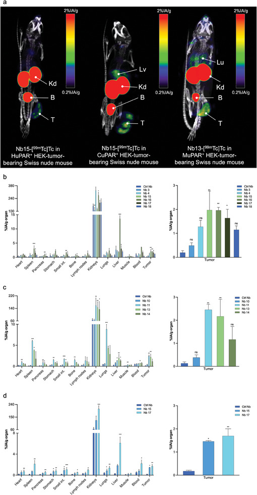
Figure 3.

a) Biodistribution of 99mTc‐labeled anti‐uPAR Nbs, in Swiss nude mice bearing subcutaneous uPAR‐transduced HEK tumors. Micro‐SPECT/CT images were obtained 1 h after intravenous injection of 99mTc‐labeled Nbs. Arrows indicate T: tumor; B: bladder; Kd: kidneys; Lv: liver, and Lu: lungs. Ex vivo biodistribution and tumor targeting of the 99mTc‐labeled anti‐uPAR Nbs and non‐targeting control Nb in Swiss nude mice subcutaneously bearing H, M or CuPAR transduced HEK tumors (b–d, respectively). Statistical analyses were performed using a Kruskal‐Wallis test followed by a Dunn's multiple comparisons test for the selection of the lead Nbs. Statistical significance was set at p < 0.05 (*p < 0.05, **p < 0.01, ***p < 0.001, ****p < 0.0001).
