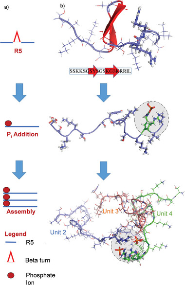
Figure 3.

Phosphate interaction and supramolecular architecture of R5 assemblies. a) Scheme depicting the tripeptide assembly mechanism as observed in the MD simulations. b) Snapshots, each after 1 µs of an MD simulation, of three MD runs. From top to bottom: R5 in water, R5 in the presence of Pi, and self‐assembly of three R5 units in the presence of Pi. The snapshots visualize the tripeptide assembly process by binding of Pi to the RRIL motifs of R5. Statistical analysis of the simulations and 31P NMR spectra of the Pi ions can be found in Figure S7 (Supporting Information).
