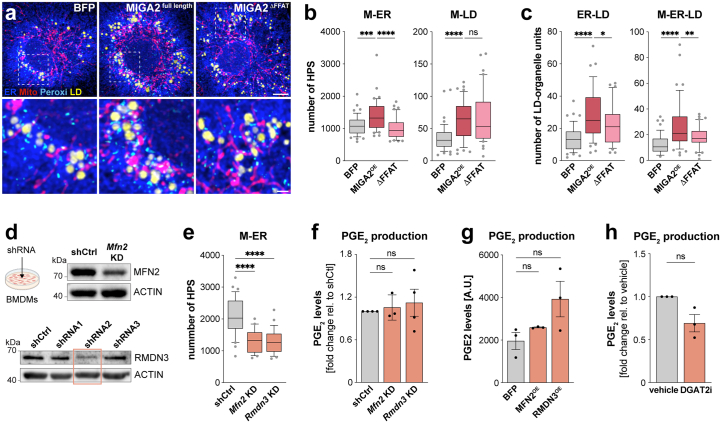
Extended Data Fig. 8. MIGA2 but not other M-ER-LD tethers contribute to macrophage PGE2 production.
a Images depicting M-ER-P-LD units in LPS/IFN-γ-activated BMDMs (24 h) expressing BFP, MIGA2 or MIGA2DFFAT. Images represent maximum intensity projections of n = 3 biolgocial replicates. Scale bars: 5 µm, 1 µm (magnification). b,c Pair-wise M-ER and M-LD high proximity sites (b) and MIGA2-regulated LD-units (c) in LPS/IFN-γ-treated (24 h) BMDMs expressing BFP, MIGA2 or MIGA2DFFAT. Data represent N = 48 (BFP), N = 48 (MIGA2) and N = 40 (MIGA2DFFAT) cells from n = 3 biologically independent replicates. Boxes represent 25th to 75th percentiles, whiskers 10th and 90th percentiles. Dots are outliers, the median is shown as a line. P values were obtained using one-way ANOVA with Dunnett’s post-hoc test. d Western Blot analysis showing knock-down (KD) efficiency of Mfn2 or Rmdn3 in BMDMs representative of n = 1 experiment. e Pairwise ER-mitochondria HPS analysis in Mfn2 KD, Rmdn3 KD or control BMDMs. Data represent N = 32;24;29 (shCtrl; Mfn2 KD; Rmdn3 KD) cells from n = 3 biological replicates. Boxes represent 25th to 75th percentiles, whiskers 10th and 90th percentiles. Dots are outliers, the median is shown as a line. P values were calculated using one-way ANOVA with Dunnett’s post hoc test. f-h PGE2 production of LPS/IFN-γ-treated BMDMs (24 h) upon Mfn2 and Rmdn3 KD (f), MFN2 and RMDN3 overexpression (g) or DGAT2 inhibition (DGAT2i) (h). Data represent n = 4 (f, Rmdn3 KD) and n = 3 (f, Mfn2 KD; g-h) biologically independent experiments. P values were calculated using two-tailed, one-sample t tests (f,h) or one-way ANOVA with Dunnett’s post-hoc test (g). Bars show mean ± SEM (f-h). Numerical P values are available in Supplementary Information Table 4 (*P ≤ 0.05, **P ≤ 0.01, ***P ≤ 0.001, ****P ≤ 0.0001, not significant (ns) P > 0.05). Source numerical data and unprocessed blots are available in source data.

