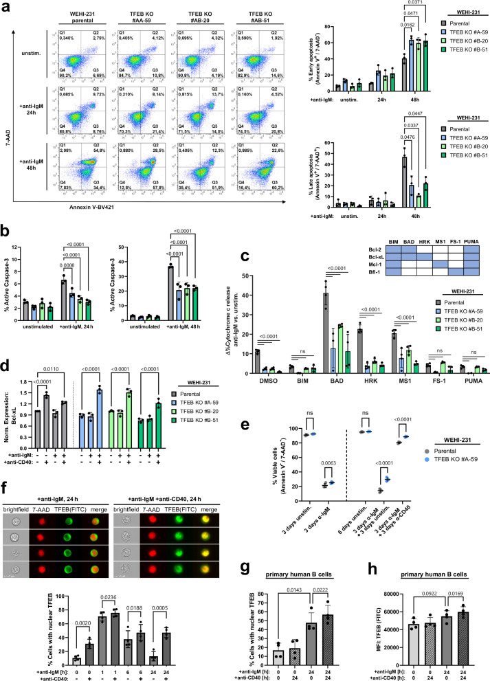
Fig. 7. TFEB balances pro-apoptotic BCR with CD40 rescue signals.
a Parental and TFEB-mutant WEHI-231 B cells were left untreated or BCR-stimulated for the indicated periods. Flow cytometric co-staining with Annexin V-BV421 and 7-AAD was used to detect early (Annexin V+/7-AAD-) and late apoptosis (Annexin V+/7-AAD+). b Flow cytometry analysis of active caspase-3 in WEHI-231 cells treated as described above. Data in (a, b) are depicted as mean percentage±SD of gated cells from n = 3 independent experiments. Significances were computed using Dunnett-corrected two-way ANOVA. c BH3 profiling of TFEB-depleted and parental WEHI-231 cells using the indicated BH3-agonistic peptides. Binding specificities towards Bcl-2 family proteins are indicated in the corresponding matrix. Cytochrome c release was monitored for resting cells or after 6 h of BCR ligation by flow cytometry. BCR-induced sensitization is presented as mean ± SD percent difference (Δ%) between n = 4 technical replicates of stimulated versus unstimulated cells. Depicted data are representative of n = 3 biological replicates. Statistical significances were calculated using Tukey-corrected one-way ANOVA. d WEHI-231 cells were incubated with the indicated combinations of anti-BCR and anti-CD40 antibodies for 6 h. Intracellular Bcl-xL expression was assessed by flow cytometry and is depicted as MFI, normalized to unstimulated parental cells. e Parental WEHI-231 and TFEB KO #A-59 cells were left untreated or stimulated, as indicated. Medium was changed on day 3, and cells were left untreated or were rescued with anti-CD40 until day 6. On day 3 and day 6, cells were incubated with Annexin V/7-AAD to access viability, as defined by a double negative staining. f WEHI-231 cells were BCR-stimulated for the indicated time periods in the presence or absence of CD40 ligation. Imaging flow cytometry-derived representative images and nuclear translocation of TFEB as percentages of cells with a TFEB/7-AAD similarity score ≥ 1. g, h Primary peripheral human CD19+ B cells were left untreated or BCR-stimulated for 24 h with or without CD40 ligation. Nuclear translocation (g) and expression (h) of TFEB was measured by imaging flow cytometry. Data shown in (d–h) is presented as mean ± SD of n = 3 (d) or n = 4 (e–h) independent experiments. Statistical significances were calculated using Tukey-corrected one-way ANOVA. Source data are provided as a Source Data file.

