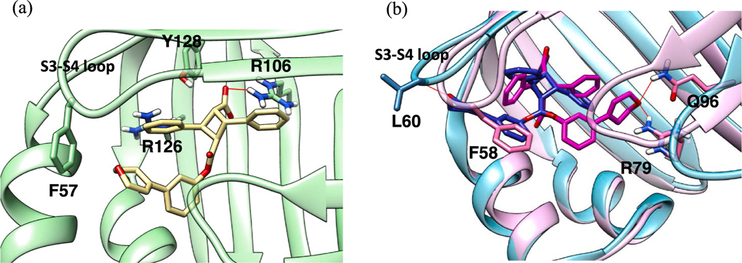
Fig. 12.
(a) Docking pose of α-21 (white) in FABP3, showing substantial dislocation of the whole molecule in the binding site, cutting off canonical interactions with Arg126 and Tyr128. (b) Docking pose of α-21 in FABP5 (protein: light blue; ligand: blue) and FABP7 (protein: pink; ligand: magenta). (For interpretation of the references to color in this figure legend, the reader is referred to the web version of this article.)

