FIG. 1.
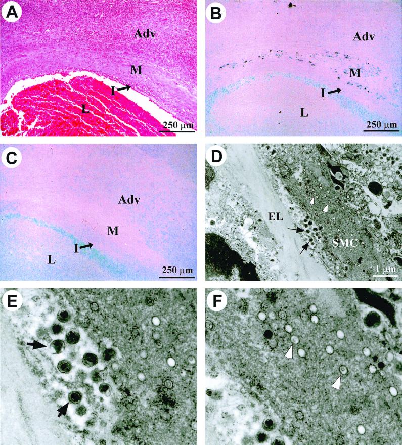
Chronic productive γHV68 infection in the arteritic media. (A through C) Serial sections from an arteritic lesion in a γHV68-infected IFN-γR−/− mouse that was sacrificed 11 weeks p.i. Adv, adventitia; M, media; I, intima; L, lumen. (A) H&E-stained section. (B) DNA in situ hybridization with a γHV68-specific DNA probe. Dark staining within the media represents specific signal. (C) DNA in situ hybridization with an MCMV-specific DNA probe. Light blue staining in the lumen with both γHV68 and MCMV DNA probes represents background staining from the glass slide. (D) Electron micrograph of the media of an arteritic lesion from an IFN-γR−/− mouse that died 6 weeks p.i. Black arrows indicate extracellular mature virions. White arrows indicate virions in the cytoplasm of a smooth muscle cell (SMC). EL, elastic lamina; SMC, smooth muscle cell. Magnification, ×11,500. (E) Enlargement of region containing extracellular virions from the electron micrograph shown in panel D. (F) Enlargement of region containing cytoplasmic virions from the electron micrograph shown in panel D.

