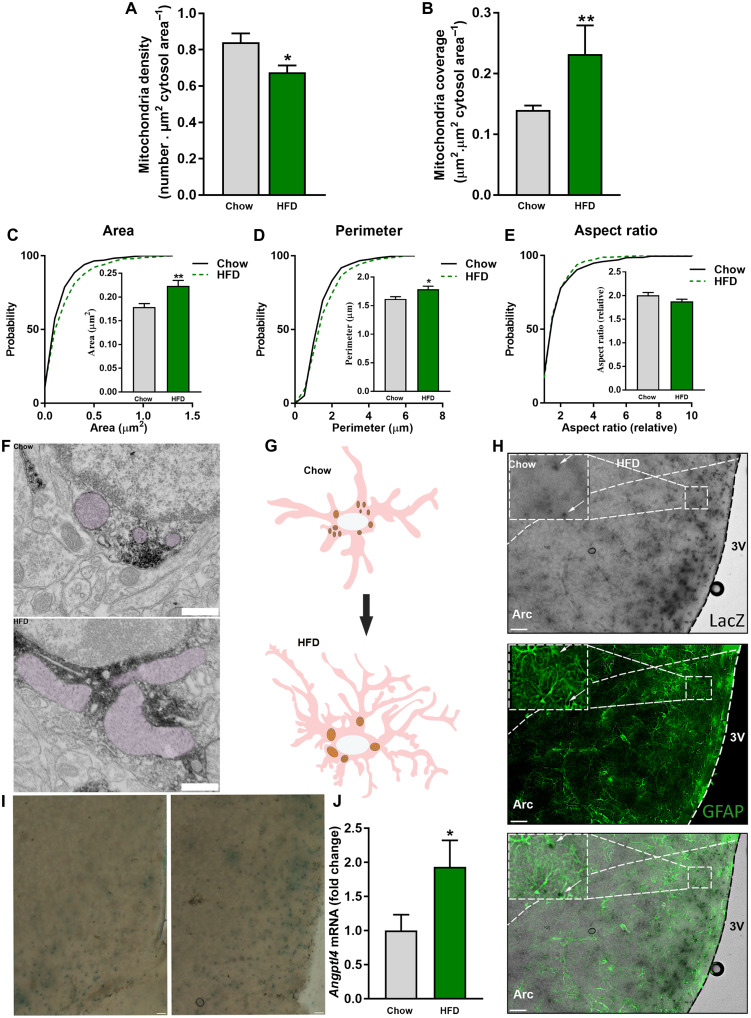
Fig. 1. HFD induces changes in astrocytic mitochondrial morphology.
Mitochondria (A) density and (B) coverage in MBH astrocytes of mice exposed to chow and HFD. Cumulative distribution and mean of mitochondria (C) area, (D) perimeter, and (E) aspect ratio of mediobasal hypothalamus (MBH) astrocytes of mice fed a chow diet and HFD (n > 350 mitochondria from >43 astrocytes from 12 and 9 mice, respectively). (F) Representative electron micrographs from MBH astrocytes of mice fed a chow diet and HFD. Scale bar, 500 nm. (G) Schematic representation of astrocytes and their mitochondria in response to HFD feeding. (H) Representative images showing colocalization of glial fibrillary acidic protein (GFAP) and ANGPTL4-LacZ. Scale bars, 100 μm. (I) Representative images showing ANGPTL4-LacZ staining in the MBH of chow- and HFD-fed mice. Scale bars, 50 μm. (J) Angptl4 mRNA levels from MBH astrocytes of mice fed a chow diet and HFD (n = 6 and 7 samples per group, respectively). Data are presented as means ± SEM. *P ≤ 0.05, and ***P ≤ 0.001 as determined by two-tailed t test or Kolmogorov-Smirnov test for analyses of cumulative distribution.

