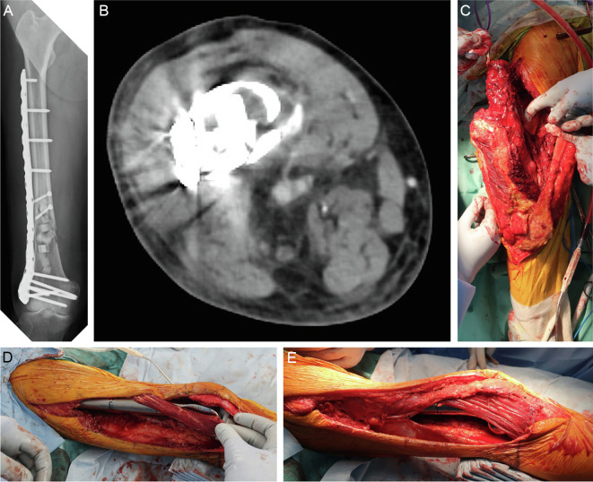
Figure 4.
Case 2: 74-year-old man. The patient experienced pain in the distal right femur, and a needle biopsy was performed based on suspicion of an enchondroma or atypical cartilaginous tumor as shown by X-ray and magnetic resonance imaging. The pathological examination also showed findings consistent with an enchondroma or atypical cartilaginous tumor, and the patient was followed up with imaging. Two years after the biopsy, the pain recurred and a pathological fracture developed. (A, B) The fracture site was identified by a lateral approach, and the tumor was curetted using a high-speed burr and cauterized with an argon beam coagulator. The resulting cavity was filled with artificial bone, and the fracture was fixed with a locking plate. Based on the pathological examination of the curettage specimen, a diagnosis of dedifferentiated chondrosarcoma was made. (C–E) Additional wide resection was performed with a 3-cm margin around the previous skin incision, and reconstruction with a megaprosthesis and sartorius muscle flap coverage was performed. Postoperative adjuvant radiation therapy of 66 Gy in 33 fractions was administered. Two months after the additional wide resection, multiple lung and bone metastases were found, and the patient died 6 months after the diagnosis of dedifferentiated chondrosarcoma.

