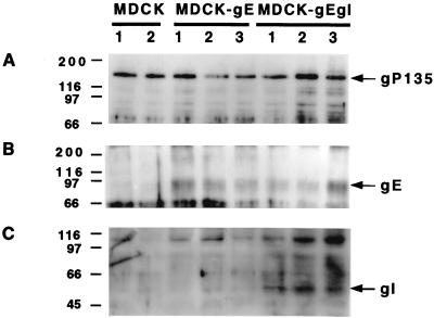
FIG. 1.
Western blot analysis of constitutively gE- or gE/gI-expressing cell lines. Two clones of MDCK control cells, three clones of MDCK-gE-expressing cells, and three clones of MDCK-gE/gI-expressing cells were grown on six-well cluster plates for 20 h and then extracted in lysis buffer. Proteins were separated by SDS–8.5% polyacrylamide gel electrophoresis, transferred to nitrocellulose membrane, and probed with anti-gP135 monoclonal antibody (A) or anti-gI monoclonal antibody (C). The same cell lysates were immunoprecipitated with anti-gE monoclonal antibody, separated by SDS–10% polyacrylamide gel electrophoresis, transferred to nitrocellulose membrane, and probed with anti-gE monoclonal antibody (B).

