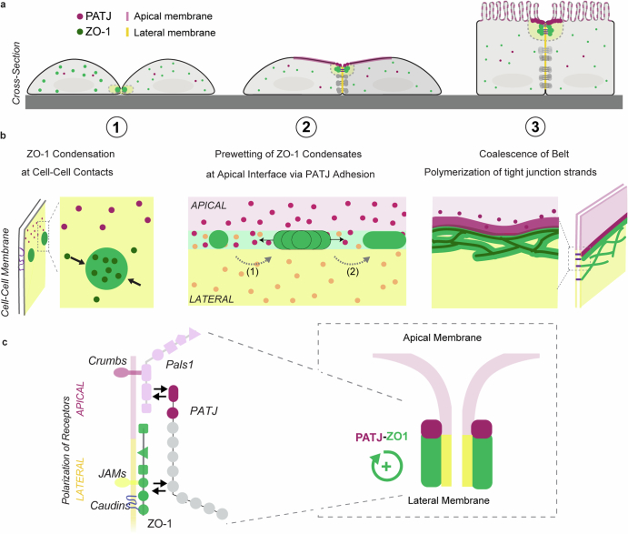
Extended Data Fig. 7. Model of tight junction belt formation by prewetting of junctional condensates along the apical membrane interface.
(a) Cross-section view of tight junction formation phases on the cellular level. (1) ZO-1 (green) condensates nucleate at cell-cell contact sites. (2) Cells polarize and nucleated ZO-1 surface condensates grow around the apical interface (magenta). (3) Cell-cell contacts mature, and the tight junction belt closes and seals the tissue. (b) Mesoscale events during tight junction belt formation. (1) Nucleation of ZO-1 condensates leads to partitioning of junction proteins. (2) Interaction of nucleated condensates with the apical interface via PATJ induces condensate growth along the interface via a prewetting transition. Growing condensates fuse into a continuous tight junction belt. (3) Polymerization of tight junction strands establishes the tight trans-epithelial barrier. (c) Molecular interactions underlying prewetting of ZO-1 condensates along the apical interface. ZO-1 surface condensates nucleate by ZO-1 binding to receptors that bind in trans at cell-cell adhesion sites (JAMs, Claudins). PATJ binds to the apical membrane via the apical complex (Pals1). Preferential interactions of membrane bound ZO-1 and PATJ mediate enrichment of the ZO-1 – PATJ complex at the apical interface. Apical interface enrichment promotes growth of ZO-1 surface condensates via prewetting.

