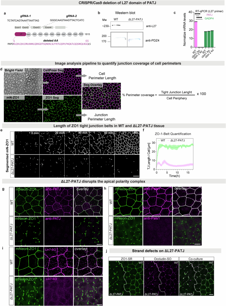
Extended Data Fig. 3. PATJ is required for tight junction formation.
(a) Deletion of L27 domain of PATJ via CRISPR/Cas9 in the background of mN-ZO-1 knock in. Guides used to remove exon 3 and sequencing confirming the deletion of the first exon and frame shift leading to an early stop codon in two independent clones. (b) Western Blot showing PATJ protein levels in WT and ∆L27-PATJ cells using anti-L27 or anti-PDZ4 domain antibodies. Antibody against the N-terminal L27 domain of PATJ, encoded by the first exon, confirmed deletion of this domain (up). Antibody against the C-terminal PDZ4 show a remaining truncated protein with a smaller size of ~ 40 kDa (low), (see Supporting Fig. 1). (c) qPCR showing the mRNA levels of PATJ in WT and two independents cell clones of ∆L27-PATJ deleted L27 domain encoded by the first exon. Data shows mean ± SD, statistical analysis was done using an unpair t test (not significant (n.s), p < 0.00001****). (d) Segmentation pipeline to quantify the coverage of cell perimeters by mN-ZO-1 condensates using CellPose for the cell perimeter on phase contrast images and local intensity thresholding and skeletonization in Fiji for mN-ZO1. (e) Segmented and skeletonized mN-ZO1 during calcium switch quantifying tight junction per cell in WT vs ∆L27-PATJ MDCK-II during 48 h. (f) Quantification of tight junction length per cell for segmented panel e, data shows mean ± SEM of n = 7 monolayers n > 50 cells. (g) Staining of PATJ (magenta) and ZO-1(green) in WT vs ∆L27-PATJ MDCK-II. Staining of Pals1 (magenta) and ZO-1(green) in WT vs ∆L27-PATJ MDCK-II. (i) Staining of Lin7 (magenta) and ZO-1(green) in WT vs ∆L27-PATJ MDCK-II. (j) Staining of Occudin (magenta) and ZO-1(green) in WT vs ∆L27-PATJ MDCK-II co-culture cells. Panels show representative images of n = 2 (b), n = 10 (d) and n = 3 biological replicates (e, f, g,h,I,j). Scale bar is 20 µm (d, e) and 10 µm for (g,h,I,j).

