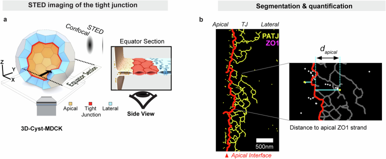
Extended Data Fig. 4. STED super-resolution imaging of the apical interface.
(a) Combining 3D tissue culture of MDCK-II cysts with STED microscopy enables super- resolution imaging of cell-cell interfaces. At the equator cell-cell interfaces are oriented parallel to the high-resolution axis (XY) of the microscope. (b) 2-color STED imaging of PATJ (yellow) and ZO-1 (magenta) in MDCK-II cysts reveal that ZO-1 forms a network structure reminiscent of tight junction strands. PATJ is enriched as clusters at the apical interface of the ZO-1 network.

