FIGURE 4.
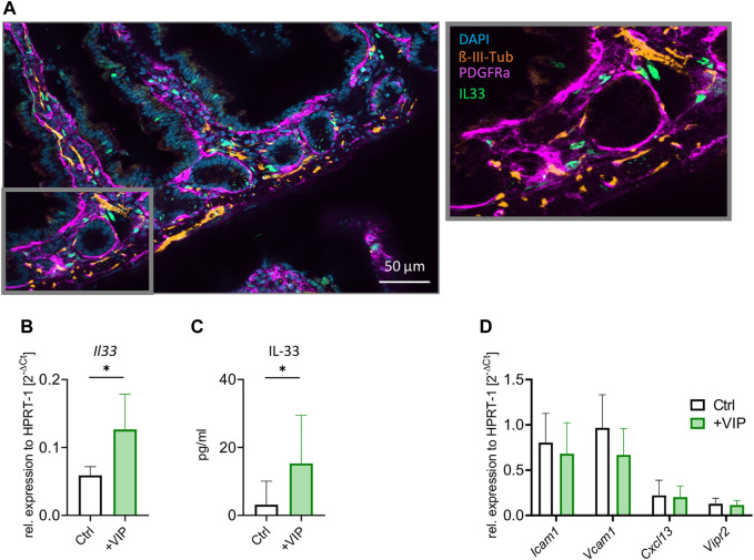
Stromal cells upregulate IL-33 in response to VIP stimulation. (A) Immunohistochemistry of the transverse section of the small intestine. Neurons stained in orange ( -III-Tubulin), stromal cells in magenta (PDGFR-α) and IL-33 antibody in green. Nuclei are stained by DAPI (blue). (B) qPCR of Il33 gene after stimulation of stromal cell culture with VIP, compared to Ctrl (unstimulated). (C) ELISA of IL-33 after stimulation of stromal cell culture with VIP, compared to Ctrl (unstimulated). (D) qPCR of Icam1, Vcam1, Cxcl13, and Vipr2 genes after stimulation of stromal cell culture with VIP, compared to Ctrl (unstimulated). (B–D): Data are representative of two experiments with three to five mice per group. Mean ± SD, Student’s t-test. *p < 0.05.

