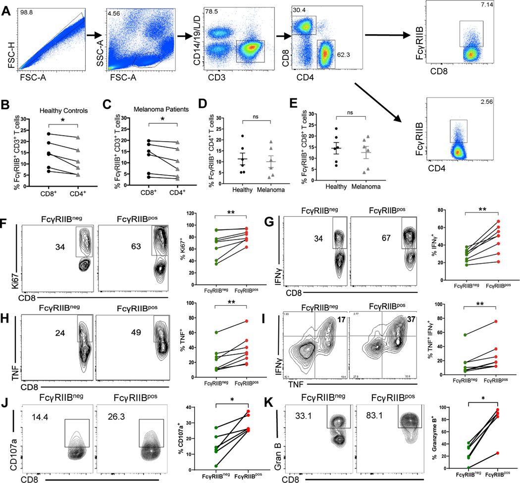
Fig. 1. FcγRIIB is expressed on CD8+ T cells from human PBMCs and is a marker of activated, cytokine-producing cells.
(A) Flow cytometry plots showing gating strategy for excluding dead cells as well as contaminating CD14+ and CD19+ cells and gating on the FcγRIIBpos population in human CD8+ and CD4+ T cell compartments of PBMCs. (B-C) Quantification comparing the frequency of FcγRIIBpos CD4+ and CD8+ T cells in (B) human healthy controls and (C) stage IV patients with melanoma. (D) Quantification comparing the frequency of FcγRIIBpos CD4+ T cells between healthy donors and stage IV melanoma patient (n=6). (E) Quantification comparing the frequency of FcγRIIBpos CD8+ T cells between healthy donors and stage IV patients with melanoma (n=6 per group). (F-K) PBMCs from healthy controls were stimulated in vitro for 5 days with anti-CD3/CD28 beads, re-stimulated with PMA/Ionomycin for 4h and stained intracellularly (n=8). Representative flow plots and quantification showing the frequency of (F) Ki-67+, of (G) IFNγ+, of (H) TNF+, of (I) TNF+ IFNγ+, of (J) CD107a+, and of (K) Granzyme B+ FcγRIIBpos and FcγRIIBneg CD8+ T cells. When comparing cell groups within the same donor, P-values were calculated using the Wilcoxon matched-pairs rank test. Mann-Whitney non-parametric, unpaired tests were used when comparing groups between donors. The error bar in summary figures denotes mean ± SEM. *P<0.05 **P<0.01, ns, not significant.

