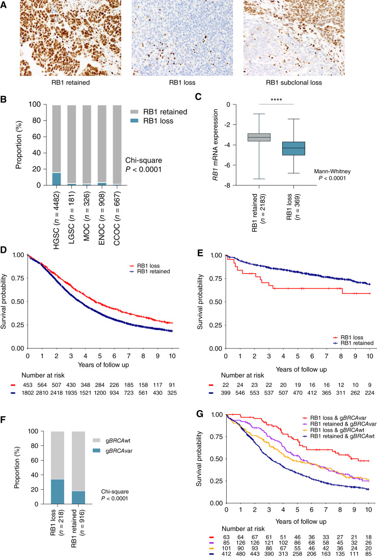
Figure 1.
Expression of RB1 and survival associations across ovarian cancer histotypes. A, Representative images of IHC detection of RB1 expression in ovarian carcinoma tissues, showing examples of the three most common expression patterns: retained, lost, and subclonal loss. B, Proportion of patients with loss or retention of RB1 protein expression in tumor samples by ovarian cancer histotypes. χ2P value reported for difference in proportions across all histotypes. CCOC, clear cell ovarian cancer; LGSC, low-grade serous carcinoma; MOC, mucinous ovarian cancer. C, Boxplots show RB1 mRNA expression (NanoString) by RB1 protein expression status; lines indicate median and whiskers show range (Mann–Whitney test P value reported). Kaplan–Meier analysis of OS in patients diagnosed with HGSC (D) and ENOC (E) stratified by tumor RB1 expression. F, Frequency of germline BRCA wild-type (gBRCAwt) and germline BRCA pathogenic variants (gBRCAvar) in patients with HGSC stratified by RB1 protein expression. χ2P value is reported. G, Kaplan–Meier estimates of overall survival in patients with HGSC by combined germline BRCA and tumor RB1 expression status.

