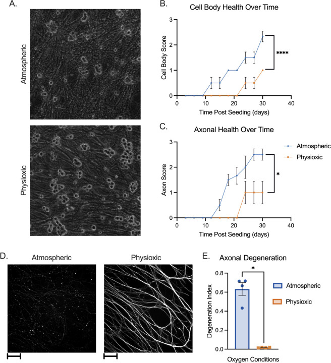
Figure 1.
Neuronal Health in Atmospheric versus Physioxic Incubation Conditions. (A-E) Primary sympathetic neurons were incubated in atmospheric (approximately 18% oxygen) or physioxic (5% oxygen) conditions. (A) Neurons were latently infected with Stayput-GFP at an MOI of 10 PFU/cell in the presence of acyclovir (ACV; 50 μM) for ten days and cultures were imaged using phase contrast to document soma morphology. (B-C) Neurons were incubated for up to 30 days post-plating. Scores representing cell body (B) and axon (C) health were recorded over time. Biological replicates from 2 independent dissections; statistical comparisons were made using 2-way ANOVA. (D-E) Neurons were plated in microfluidic chambers. 10 days post-plating, cultures were fixed and stained for neuronal marker Beta III tubulin (white) (D). Scale bar 100 μm. Axonal degeneration was analyzed using these images (E). Biological replicates from 4 independent experiments; statistical comparisons were made using unpaired non-normal t-test. Individual biological replicates along with the means and SEMs are represented. * P < 0.05; ** P < 0.01.

