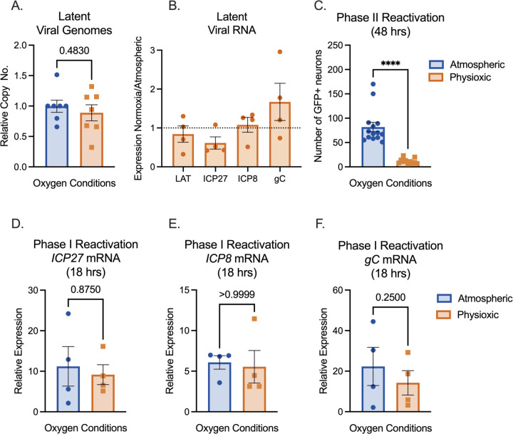
Figure 2.
Physioxic incubation conditions reduce HSV-1 reactivation. (A-F) Primary neurons were latently infected with Stayput-GFP at an MOI of 10 PFU/cell in the presence of acyclovir (ACV; 50 μM) for six days and then reactivated two days after the removal of acyclovir with LY294002 (20 μM). Quantification of relative latent viral DNA load (A) and LAT expression (B) at 8 days post-infection. C) Quantification of the number of GFP-positive neurons at 48 hours post-stimulus. D-F) Relative viral gene expression at 18 hours post-stimulus compared to latent samples quantified by RT-qPCR for immediate early viral gene ICP27 (D), early viral gene ICP8 (E), or late gene gC (F) normalized to cellular control mGAPDH. Biological replicates from 4 independent dissections; statistical comparisons were made using paired t-tests. Individual biological replicates along with the means and SEMs are represented. * P < 0.05; ** P < 0.01.

