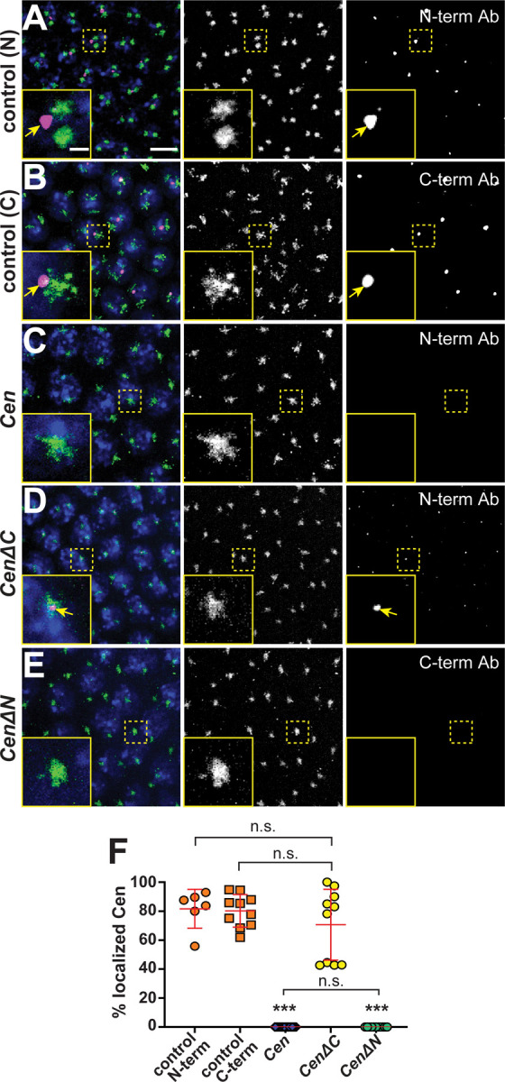
Figure 3. he N-terminal fragment is necessary and sufficient for Cen protein localization to the centrosome.

T Maximum-intensity projections of NC 13 interphase embryos expressing GFP-Cnn (green) labeled with anti-Cen antibodies (magenta) and DAPI (blue nuclei). Control embryos labeled with (A) anti-Cen N-terminal or (B) C-terminal antibodies (Ab) show Cen localized at centrosomes (arrows). (C) Cen protein is not detected in null mutants. (D) The N-terminal fragment (CenΔC) is sufficient to direct Cen to the centrosome (arrows), while the C-terminal fragment (CenΔN; (E)) is not. Both transgenes are expressed in the Cen null background. (F) The percentage of Cen protein signals overlapping with centrosomes (0 μm from Cnn surface). Each dot represents a measurement from N= 6 control (N-terminal Cen Ab), 10 control (C-terminal Cen Ab), 23 Cen null (N-terminal Cen Ab), 10 CenΔC (N-terminal Cen Ab), and 11 CenΔN embryos (C-terminal Cen Ab). Significance was determined by Kruskal-Wallis test followed by Dunn’s multiple comparison test with n.s., not significant and ***, P<0.001. Scale bars: 5μm; 1μm (insets).
