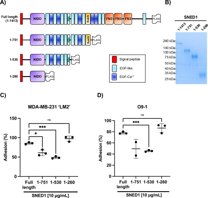
Figure 2. The N-terminal region of SNED1 mediates cell adhesion.
(A) Schematic showing FLAG-tagged full-length SNED1 and the different truncated forms of SNED1 used in this study: SNED11–751 encompasses the N-terminal region until the sushi domain; SNED11–530 encompasses the N-terminal region until the follistatin domain; SNED11–260 encompasses the N-terminal region until the NIDO domain.
(B) Coomassie-stained gel showing the purity of purified full-length and truncated forms of SNED1.
(C-D) Adhesion of MDA-MB-231 ‘LM2’ breast cancer cells (C) and O9–1 neural crest cells (D) to full-length and truncated forms of SNED1. Data is represented as mean ± SD from at least two biological experiments. Welch and Brown-Forsythe one-way ANOVA with Dunnett’s T3 correction for multiple comparisons was performed to determine statistical significance. ns: non-significant, *p<0.05, *** p<0.001.

