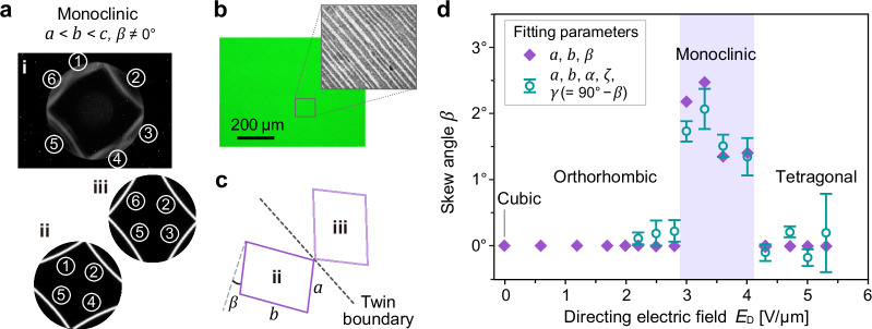
Fig. 5. Twinned crystal with monoclinic symmetry.
A twinned monoclinic crystal is formed by cooling a tetragonal single crystal (from 31.6 °C to 30.7 °C) with field maintained at 3.6 V/μm. a Kossel diffraction pattern (i) showing a total of six rings (or, arcs due to limited field of view), which can be decomposed into two sets of Kossel rings (ii, iii). The two sets share rings ② and ⑤, and each set represents one of the twins. Both patterns (ii, iii) appear as a sheared version of the Kossel pattern observed in an orthorhombic crystal (cf. Fig. 4d(ii)), suggesting the presence of monoclinic crystals. b Optical micrographs of the twinned monoclinic crystal. Contrast and brightness of the grayscale micrograph in the inset are adjusted to show the striated texture due to crystal twinning. Surface-alignment (rubbing) direction is from left to right (a, b). c Schematic depicting the unit-cell orientations of monoclinic twins on opposite sides of twin boundary (dashed line), and definition of skew angle β. d Skew angle β of monoclinic BPLC as a function of the directing field strength (ED) during REDA. At ED between ~3 and ~4 V/μm2, monoclinic crystals (β ≠ 0) can be formed through direct cooling from a tetragonal single crystal. Purple diamonds represent fits with a, b, and β as free parameters. Green open circles represent a more rigorous analysis where the angles between all three basis vectors (a, b, and c) are treated as independent fitting parameters, α, ζ, γ (= 90° − β) (details in Supplementary Note 2). Error bars represent the standard deviation.

