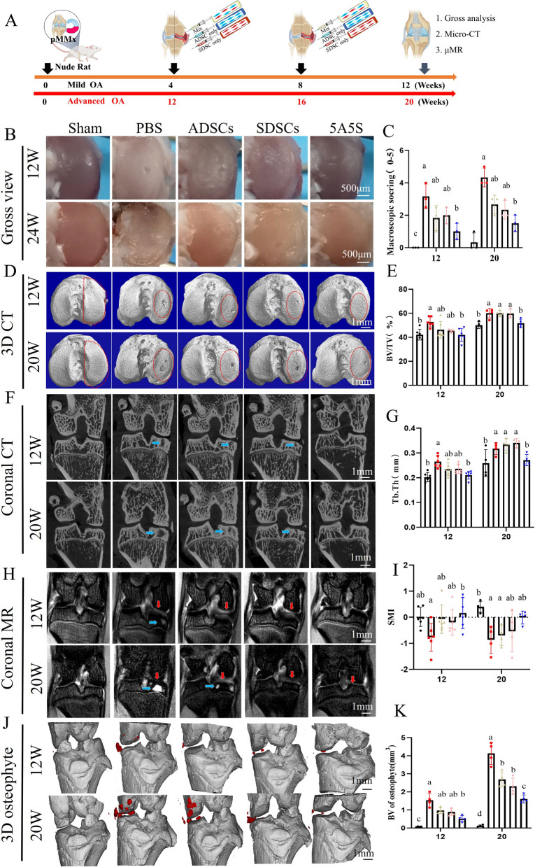
Fig. 3.
A combination of ADSCs and SDSCs in equal amounts improved the subchondral bone and osteophytes changes in early and advanced OA. A schematic illustration of intra-injection of ADSCs and SDSCs for OA therapy. B the gross view of the tibial articular surface and macroscopic scores (C) (n = 3). D 3D reconstruction of subchondral bone, E BV/TV, G Tb.Th and I SMI for medial subchondral bone (n=6 or 4). F coronal view of subchondral bone. H subchondral bone cyst (blue arrow) and cartilage edema (red arrow) by MRI images. J 3D reconstruction (marked in red) and K bone volume of the osteophytes around the knee joint (n = 4). All data are shown as the mean ± SD

