. 2024 May 20;10(2):e004036. doi: 10.1136/rmdopen-2023-004036

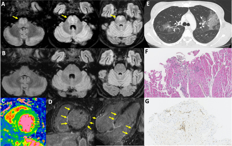
Figure 2. Organ involvement in severe enteroviral infections. (A) Brain MRI taken in June 2022 showing centro-pontine fluid-attenuated inversion recovery (FLAIR) hyperintensity without enhancement for the patient presented in figure 3; (B) brain MRI showing complete regression of abnormalities in September 2022 (same patient); (C) diffuse myocardial oedema (MRI, T2 mapping); (D) septal, basal lateral and lateral medial enhancement (MRI, late gadolinium enhancement); (E) multifocal ground glass opacities; (F and G) lymphocytic myocarditis with interstitial T cell (G:CD3+ cells) foci, dystrophic myocytes, no giant cell or eosinophilic infiltration, no myocardial necrosis.

Figure 2