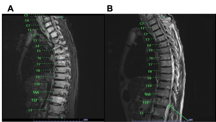
Figure 2. (A) A STIR MRI sequence of the lower cervical and thoracic spine. The radiologist noted diffuse marrow heterogeneity specifically in C7, T5, and T12 vertebral bodies. (B) A T2 weighted MRI demonstrating a sliver of epidural soft tissue along the right aspect of T12, noted with an arrow. No significant cord compression was observed, however.
STIR: Short tau inversion recovery

