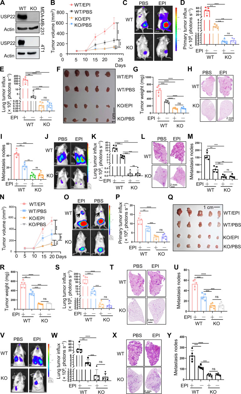
Fig. 2. EPI increases the expression of ATGL to induce the lipolysis of breast cancer through USP22.
(A) Validation of USP22 protein expression in WT and USP22 KO MDA-MB-231 cells (top) or 4T1 cells (bottom). (B to I) A total of 1 × 10⁶ WT or USP22 KO MDA-MB-231 cells were injected into the mammary fat pad of immune compromised NYG mice and treated with PBS or EPI (2 mg/kg) per day for 7 days. Tumor volume (B), bioluminescence activity [(C) and (D)], and tumor weight [(F) and (G)] were measured. Lung metastases were measured by luminol fluorescence [(C) and (E)] and H&E staining of their lung sections [(H) and (I)]. N = 5 each group. (J to M) A total of 0.5 × 10⁶ WT or USP22 KO MDA-MB-231 cells were injected via the tail vein into NYG mice. Lung metastases were measured by luminol fluorescence [(J) and (K)] and H&E staining [(L) and (M)]. (N to U) A total of 1 × 10⁵ WT or USP22 KO 4T1 cells were injected into the mammary fat pad of BALB/c mice and treated with PBS or EPI (2 mg/kg) per day for 7 days. Tumor volume (N), bioluminescence activity [(O) and (P)], and tumor weight [(Q) and (R)] were measured. Lung metastases were measured by luminol fluorescence [(O) and (S)] and H&E staining of their lung sections [(T) and (U)]. N = 5 each group. (V to Y) A total of 0.5 × 10⁵ WT or USP22 KO 4T1 cells were injected via the tail vein into BALB/c mice. Lung metastases were measured by luminol fluorescence [(V) and (W)] and H&E staining [(X) and (Y)]. Statistical significance was determined by one-way ANOVA test or unpaired Student’s t test. Data are expressed as means ± SD of five independent experiments. **P < 0.01; ***P < 0.001; ****P < 0.0001; ns, no statistical significance.

