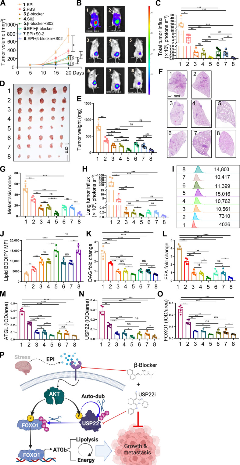
Fig. 7. Synergistic effects of USP22 and β receptor inhibitors on tumors.
(A to E) 4T1 TNBC cells were injected into the mammary fat pad of BALB/c mice (N = 5 each group). Tumor volume (A), bioluminescence activity [(B) and (C)], and tumor weight [(D) and (E)] were measured. (F to H) The metastases of the lung were measured by luminol fluorescence [(B) and (H)] and H&E staining [(F) and (G)]. Lung tissue sections were analyzed by H&E staining [(F) and (G)]. Representative lung metastasis (F) and quantifications of five mice each group (G) are shown. (I to L) Lipid analysis of syngeneic tumors from (D) by lipid BODIPY [(I) and (J)], DAG (K), and FFAs (L). MFI, mean fluorescence intensity. (M to O) IHC analysis of USP22, FOXO1, and ATGL expression levels in syngeneic tumor tissue sections from (D). Quantification data of all five mice each group [(M) to (O)] are shown. (P) Model of targeting chronic stress–mediated cancer metastasis and lipolysis by USP22. Statistical significance was determined by one-way ANOVA test or unpaired Student’s t test. Data are expressed as means ± SD of five independent experiments. *P < 0.05; **P < 0.01; ***P < 0.001; ****P < 0.0001; ns, no statistical significance.

