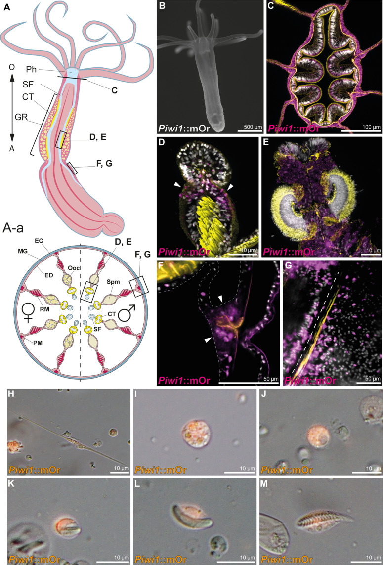
Fig. 1. Piwi1 reporter expression in somatic cell types.
(A) Whole-body and transversal section schematic indicating tissues and location of piwi1::mOr-expressing cells. (A) O, oral pole; A, aboral pole; Ph, pharynx; SF, septal filament; CT, ciliated tract; GR, gonadal region. (A-a) EC, ectoderm; MG, mesoglea; ED, endomesoderm; PM, parietal muscle; RM, retractor muscle; Ooc, oocytes; Spm, spermaries. (B) Juvenile piwi1::mOr polyp. mOr is displayed as white. (C) Section through a pharynx. (D) Juvenile mesentery. Arrows mark piwi1+ cells accumulating at the gonad primordium. (E) Ciliated tract of an adult mesentery. (F) Piwi1+ cells concentrated at the mesentery base (arrowheads), surrounding the parietal muscle (orange). (G) Optical section along the mesentery base showing piwi1+ cells along the parietal muscle; the dotted line indicates the mesentery base. (H to M) Piwi1::mOr-positive cell types. (H) Neuron. (I) Gland cell. (J) Oocyte. (K) Immature nematocyte. (L) Mature nematocyte. (M) Spirocyte. For all fluorescent pictures, 4′,6-diamidino-2-phenylindole (DAPI) is displayed as white, and phalloidin is displayed as yellow.

