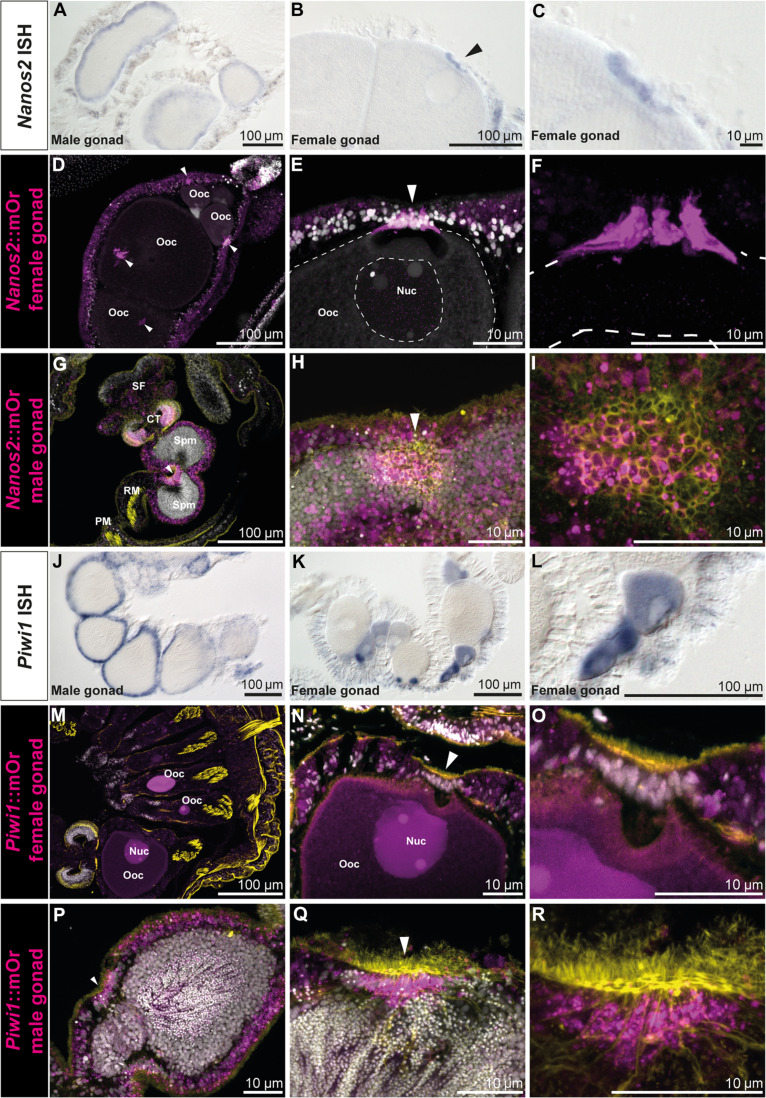
Fig. 3. Nanos2 and piwi1 mRNA and reporter expression in gametes and the gonadal region.
(A) Nanos2 expression in male mesenteries. (B) Nanos2 expression in female mesenteries. Nanos2+ trophocytes are marked by an arrow. (C) Detailed picture of (B). (D to I) Nanos2::mOr expression in adult gonads. (D) Female gonad showing reporter-expressing trophonemata located closely to the germinal vesicle of each oocyte (arrowheads). (E) Nanos2::mOr-expressing trophonema (arrowhead). Nuc, nucleus of the oocyte. (F) Detailed picture of (E) (DAPI not displayed). (G) Male gonad, with mOr expressing in the gastrodermis around the spermaries and the bordering tissue of the ciliated tract. The ciliated plug is indicated by an arrowhead. (H) Detailed picture of a ciliated plug (arrowhead) in the gastrodermis surrounding the sperm chambers. Cells composing the plug can be identified by increased mOr expression and phalloidin incorporation (yellow). (I) Detailed picture of (H) (DAPI not displayed). (J) Piwi1::mOr expression in male mesenteries. (K) Piwi1::mOr expression in female mesenteries. (L) Detailed picture of the mOr+ maturing oocytes in (K). (M to R) Piwi1::mOr expression in adult gonads. (M) Female gonad. (N) Oocyte with the mOr− trophonema indicated by an arrowhead. (O) Detailed picture of (N). (P) Male gonad expressing piwi1::mOr in the endomesoderm surrounding the sperm chambers. The ciliated plug is indicated by an arrowhead. (Q) A ciliated plug (arrowhead) in the gastrodermis surrounding the sperm chambers. Cells composing the plug can be identified by increased mOr expression and phalloidin incorporation (yellow). (R) Detailed picture of (Q) (DAPI not displayed). For all fluorescent pictures, mOr is displayed as magenta, DAPI is displayed as white, and phalloidin is displayed as yellow.

