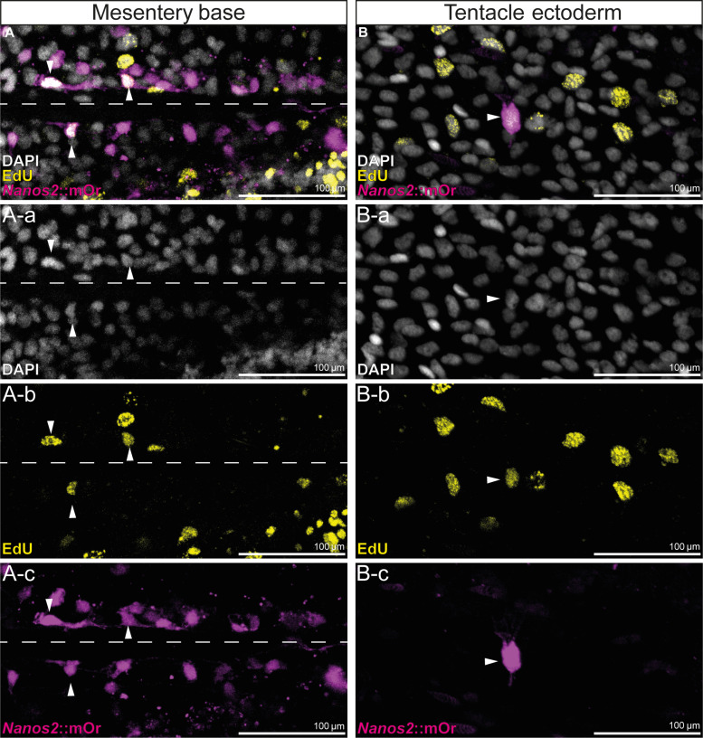
Fig. 4. EdU incorporation of nanos2::mOr-expressing cells.
(A) A-a to A-c: Fluorescent pictures of the endoderm at the base of the mesentery. The attachment site of the mesentery is indicated by a dotted line. mOr/EdU+ cells are indicated by arrowheads. (B) B-a to B-c: Fluorescent pictures of the tentacle ectoderm. mOr/EdU+ cell is indicated by an arrowhead.

