Figure 4.
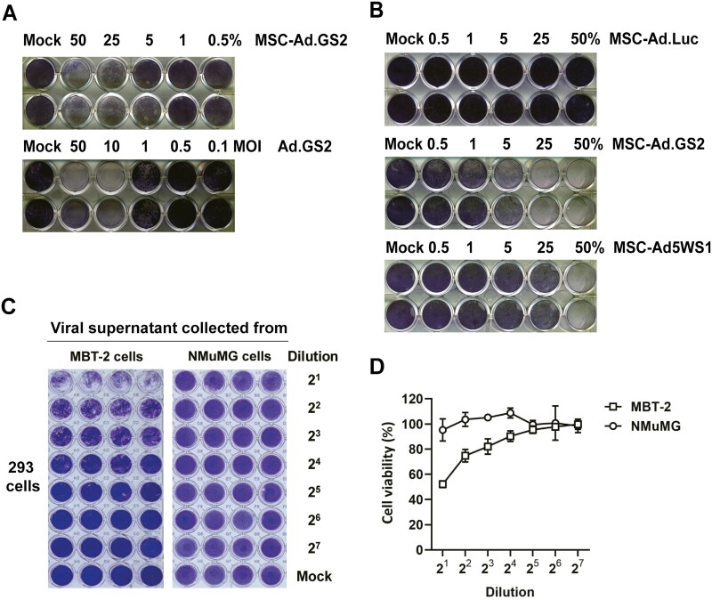
MSC-Ad.GS2 exhibits potent cytolytic effects on MBT-2 cells. (A) MSC-Ad.GS2 is superior to Ad.GS2 in lysing MBT-2 cells. MSCs were infected with Ad.GS2 at an MOI of 100. After 24 h, Ad.GS2-infected MSCs (MSC-Ad.GS2) were detached and cocultured with MBT-2 cells in ratios ranging from 0.5% to 50%. Direct cytolytic effect of Ad.GS2 on MBT-2 cells was estimated in parallel by direct infection with Ad.GS2 at different MOI. After 7 days, MBT-2 cells were examined for CPE by crystal violet staining. (B) MSC-Ad.GS2 exerts higher cytolytic effects on MBT-2 cells than MSC-Ad5WS1. MSCs were infected with Ad.GS2, Ad5WS1, or Ad.Luc at an MOI of 100, followed by the same procedure as described in A. (C, D) Ad.GS2 replicates in MBT-2 cells, but not in normal NMuMG cells. MBT-2 and NMuMG cells (2 × 10⁵) were infected with Ad.GS2 at an MOI of 200 on day 0. Culture supernatants and freeze-thaw cell lysates from Ad.GS2-infected cells were collected and pooled (1 mL) to serve as crude viral supernatants on day 5. Then, 150 μL of serially diluted viral supernatants (600 μL virus supernatant plus 600 μL culture medium as 2 × dilution) were used to infect 3 × 10⁴ 293 cells cultured in 96-well plates for detecting infectious viruses. After 6 days, CPE was detected by crystal violet staining (C). The wells were scanned, and crystal violet staining was quantified to determine cell survival using the ImageJ software (D). Values shown are the percentages of cell survival, with the levels in the mock-infected cells arbitrarily set to 100.

