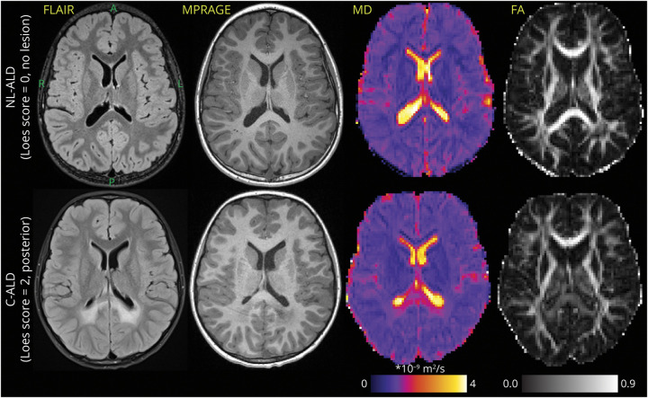
Figure 2. Representative Examples of FLAIR and MPRAGE Scans With MD and FA Maps in 2 Similar-Aged Boys With ALD.
Top row: patient without cerebral demyelinating disease. Bottom row: patient with posterior pattern C-ALD affecting the CC splenium and adjacent parietal-occipital WM (Loes = 2). Axial slices follow the orientation noted for the left top image (A = anterior, P = posterior, R = right, L = left). The interpatient slice location was matched as closely as possible across different MRI contrasts. ALD = adrenoleukodystrophy; C-ALD = cerebral adrenoleukodystrophy; FA = fractional anisotropy; FLAIR = fluid-attenuated inversion recovery; MD = mean diffusivity; MPRAGE = magnetization-prepared rapid gradient echo; WM = white matter.

