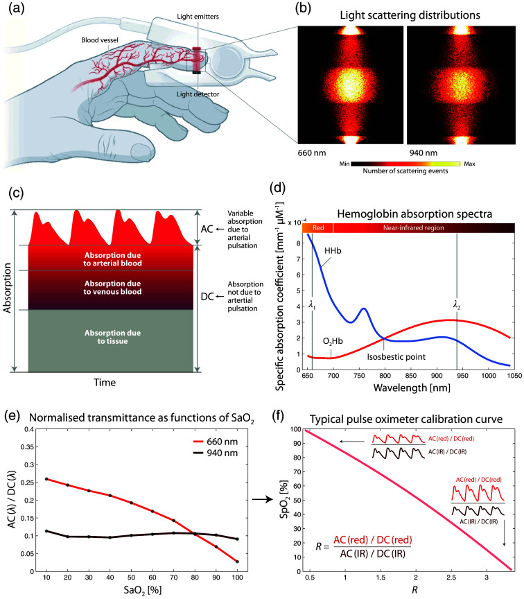
Fig. 1.
Principle of operation of PO. (a) Representative example of a finger clip of a PO, comprising light detectors and light emitters. Reprinted and modified from Kozlov,60 with permission from the publisher. (b) Light scattering distributions in the finger tissue for two wavelengths typically used in PO (red: 660 nm, infrared [IR]: 940 nm). Reprinted and modified from Chatterjee and Kyriacou.61 (c) Components of light absorption in PO (visualization redrawn from Moyle62). (d) Hb absorption spectra (data source: Biomedical Optics Research Laboratory, UCL, UK). The isosbestic point and two typical PO wavelengths are added. (e) Normalized light transmittance for two wavelengths as functions of (data: simulation by Chatterjee and Kyriacou61). A typical PO calibration curve (data source:62) with visualization of the amplitudes of the blood volume pulsation in the two wavelength ranges (red and IR). AC: alternating current (variable signal component), DC: direct current (constant signal component), : oxyhemoglobin, HHb: deoxyhemoglobin, : arterial blood oxygenation, : peripheral arterial hemoglobin saturation.

