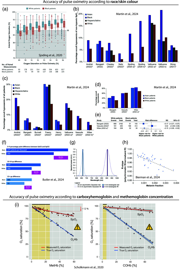
Fig. 9.
Problems with the accuracy of PO. (a) Bias in readings in Black patients compared to White patients according to the study of Sjoding et al.45 The values are plotted in comparison to the values measured in parallel. Ideally, both values should be identical. A larger discrepancy can be seen in the Black patients compared to White patients. Reprinted from Sjoding et al.,45 with permission from the publisher. (b)–(e) Results of the systematic review of Martin et al.239 (b) Frequency (in %) of occult hypoxemia in paired measurements among readings/patients with non-hypoxemic . (c) Frequency (in %) of occult hypoxemia in paired measurements out of all patients. (d) Clinical studies with respective percentages of readings with occult hypoxemia out of readings/patients with true hypoxemia. In panels (b)–(e), it can be seen that Blacks have generally a higher value, indicating a PO bias. (e) Forest plot visualizing the risk difference for Black and White patients of occult hypoxemia compared to true hypoxemia. Panels (b)–(e) reprinted from Martin et al.,239 with permission from the publisher. (f) Study results of Butler et al.240 Reprinted from Butler et al.,240 with permission from the publisher. (g), (h) Results of the simulation study of Bierman et al. showing that the spectral bandwidth (visualized in panel (g) as either Gaussian functions of LED or discrete values of laser diodes) affect how strong a PO is biased by melanin present in the tissue (h). Reprinted from Bierman et al.,241 with permission from the publisher. (i) Effect of the MetHb and COHb concentration in the blood on the accuracy of determined with a PO; reprinted from Scholkmann et al.,242 with permission from the publisher. MetHb: methemoglobin, COHb: carboxyhemoglobin, : oxyhemoglobin, : arterial hemoglobin saturation, : peripheral arterial hemoglobin saturation.

