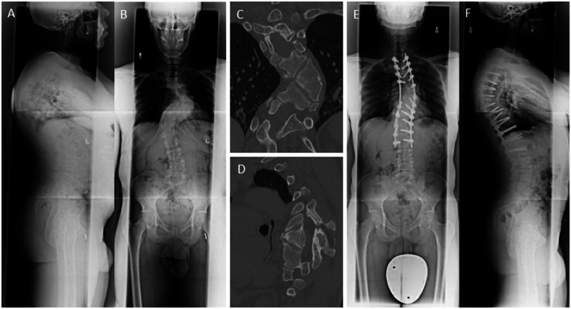
Fig. 1. Case 1, a 48-year-old healthy man with a chief complaint of mid-thoracic and low back pain with a history of congenital scoliosis diagnosed at 12 years of age.
A Anterior-posterior (AP) and (B) lateral standing radiographs demonstrated a kyphoscoliotic deformity localized at the main thoracic level, measuring 84° on Cobb angle coronally. Selective computed tomography (C) coronal and (D) sagittal images revealed unsegmented hemivertebrae at the T7 and T8 levels. E, F The patient underwent a T7 vertebral column resection (VCR) and T2-L2 posterior spinal fusion. The procedure was complicated by the intraoperative bilateral loss of motor evoked potentials. Postoperatively, the patient woke with complete loss of power in bilateral lower extremities and preservation of sensation. He was diagnosed with a T9 incomplete SCI and treatment with Riluzole was begun immediately. On the day of discharge, his lower limb power improved to 3/5 motor power bilaterally in all muscle groups. He made a significant recovery in rehabilitation and at 6 months follow up he was ambulatory with grade 5/5 motor strength in bilateral lower extremities.

