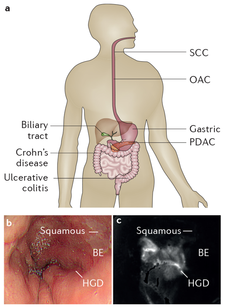
Figure 1 |. Diseases throughout the digestive tract are being evaluated in vivo with new endoscopic imaging technologies that identify molecular changes.

a | Key areas and conditions that are being examined by emerging endoscopic technology. b | White-light endoscopic image shows squamous and BE but no distinguishing features for HGD from human oesophagus in vivo. c | After topical administration of molecular probe, ratio image collected with multimodal endoscope shows region of increased intensity (arrow) from HGD5. BE, Barrett oesophagus; HGD, high-grade dysplasia; OAC, oesophageal adenocarcinoma; PDAC, pancreatic adenocarcinoma; SCC, squamous cell carcinoma.
