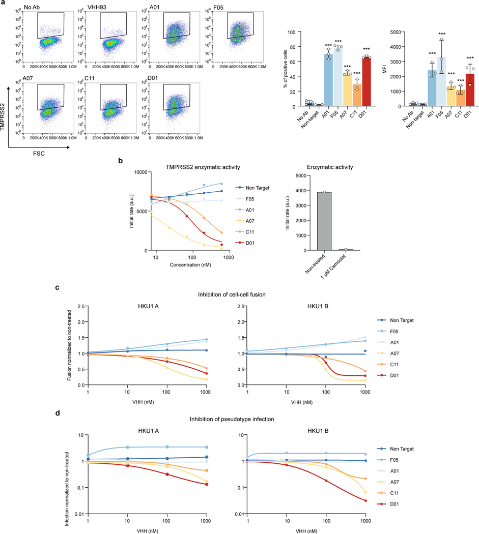
Extended Data figure 7. Characterization of anti-TMPRSS2 VHHs. a. Binding of VHHs on TMPRSS2-expressing cells.
293T cells were transfected with TMPRSS2 S441A. Binding of the indicated VHHs (0.5 μM) was assessed by flow cytometry. Left: representative dot plots. Right: Quantification of the percentage of positive cells and MFI (Median Fluorescent Intensity of positive cells). Data are mean ± SD of three independent experiments b. Effect of VHHs or Camostat on TMPRSS2 enzymatic activity. Recombinant soluble TMPRSS2 was incubated with the indicated VHHs at different concentrations or with Camostat (1 μM). The initial rate of enzymatic activity, measured with a fluorescent substrate is plotted. One representative experiment of 3 is shown. c. Effect of VHHs on HKU1A or HKU1B cell-cell fusion. 293T GFP-Split cells were cotransfected with TMPRSS2 and HKU1 spike in the presence of indicated amounts of VHH. Fusion was quantified by measuring the GFP area after 20 h. Data were normalized to the non-treated condition for each spike. d. Effect of VHHs on HKU1A or HKU1B pseudovirus infection. 293T cells transfected with S441A TMPRSS2 were incubated with the indicated amounts of VHH for 2 h and infected by Luc-encoding pseudovirus. Luminescence was read 48 h post infection. Data were normalized to the non-treated condition for each virus. Data are from one experiment representative of 3. Statistical analysis: c: One Way ANOVA with Dunnett’s multiple comparisons compared to cells stained with a non-target antibody (VHH93). d: One Way ANOVA on log-transformed data with Dunnett’s multiple comparisons compared to cells stained with a non-target antibody (VHH93).

