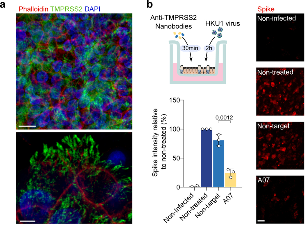
Figure 5. Live HKU1 virus infection of human bronchial epithelial (HBE) cells.
a. TMPRSS2 staining of HBE cells. Red: Phalloidin, Blue: DAPI, Green: TMPRSS2 stained with VHH A01-Fc. Scale bars: top, 10 μm; bottom, 5 μm. Images are representative of 3 independent experiments. b. Effect of the anti-TMPRSS2 VHH (A07) on HKU1 infection. The experimental design is represented. Infected cells were visualized with an anti-spike antibody and scored. Representative images of spike staining 48 h post-infection are shown. Spike pixel intensity in 5 random fields per experiment was measured and normalized to the intensity in the infected but non-treated condition. Data are mean ± SD of 3 independent experiments for infected conditions, and mean of 2 for uninfected condition. Scale bar: 20 μm. Statistical analysis: Two-sided unpaired t-test compared to non-target VHH.

