. 2024 Aug 13;12(8):e009492. doi: 10.1136/jitc-2024-009492

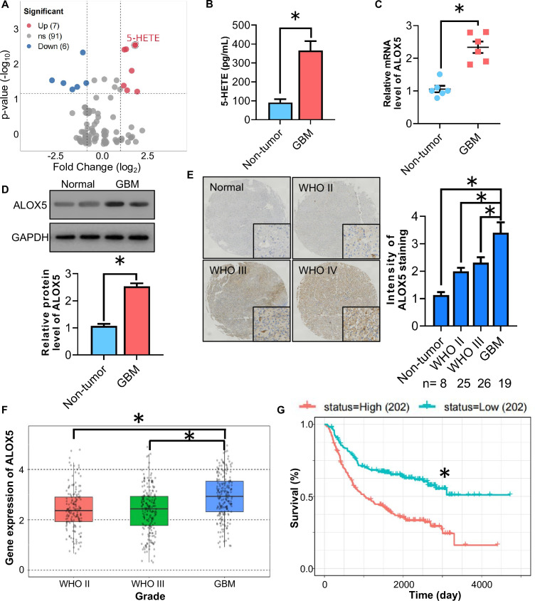
Figure 1. ALOX5/5-HETE axis is upregulated in human GBM specimen. (A) Volcano plot of oxylipin between human GBM and non-tumor brain tissues detected by targeted UHPLC-MS/MS analysis (n=10). (B) 5-HETE levels in human GBM and non-tumor brain tissues detected by targeted LC-MS/MS analysis (n=6). (C–D) Human GBM tissues and normal brain tissues were used. (C) Relative mRNA level of ALOX5 normalized with GAPDH in human GBM tissues and non-tumor brain tissues. The mRNA level is expressed as fold change over non-tumor brain tissues (n=6). (D) Immunoblotting analysis of ALOX5 and GAPDH in human GBM tissues and non-tumor brain tissues. Representative immunoblot images are shown. The bar chart is a relative expression level of ALOX5 normalized with GAPDH (n=8). (E) Immunohistochemistry staining of ALOX5 in human GBM tissue microarray. All data are represented as the mean±SEM *p<0.05 (Student’s t-test). (F) CGGA analysis of ALOX5 mRNA level in open access data sets. (G) Comparison of survival prognosis between ALOX5 high and ALOX5 low *p<0.05 (log-rank test). ALOX5, arachidonate lipoxygenase 5; GBM, glioblastoma; mRNA, messenger RNA; UHPLC-MS/MS, Ultra-high performance liquid chromatography-MS/MS; 5-HETE, 5-hydroxyeicosatetraenoic acid.

Figure 1