. 2024 Aug 13;10(3):e004201. doi: 10.1136/rmdopen-2024-004201

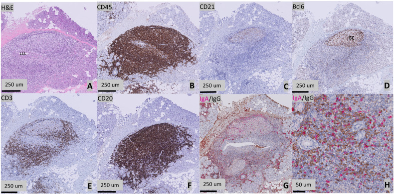
Figure 1. Histopathological features of a parotid gland of a 14-year-old patient with Sjögren’s disease (SjD). (A) H&E staining. Presence of a periductal infiltrate of ≥50 lymphocytes (focus) surrounding a striated duct with ductal hyperplasia (lymphoepithelial lesion, LEL). (B) CD45 staining showing lymphocytic infiltration. (C) CD21 staining showing follicular dendritic cell (FDC) networks. (D) Presence of a germinal centre, defined as a cluster of ≥5 adjacent Bcl6+ cells. (E) CD3 staining showing T-cell infiltration. (F) CD20 staining showing B-cell infiltration. (G) Presence of a plasma cell shift, as shown by a relative increase in the number of immunoglobulin G (IgG) (brown) plasma cells compared with the IgA (pink) plasma cells. (H) High magnification image of the IgA/IgG staining.

Figure 1