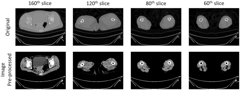
Figure 3. Example image of pre-processing on CT scans.
The outcomes of pre-processing techniques applied in image processing to enhance the accuracy of the automatic segmentation model. This step involved augmenting the contrast within CT images to distinctly delineate tissues of muscle, fat and bone. The process adjusted the intensity range of the original CT image from −57 to 164, targeting the enhancement of differentiation among various muscle tissues within the scans. Additionally, a gamma value of two was utilized to modify the contrast, thereby improving the clarity of the images. The pre-processing procedure intensified contrast and reduced metal artifacts of CT scans.

