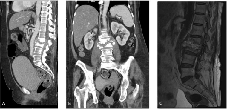
Figure 1.
(A) Sagittal section of a CT scan depicting L2–L3 discitis-osteomyelitis. (B) Coronal section of a CT scan depicting L2–L3 discitis-osteomyelitis. (C) Sagittal MRI of the lumbar spine demonstrating severe L2–L3 discitis and L2–L3 osteomyelitis with a large anterior epidural abscess causing severe spinal stenosis at the L2 and L3 levels.

