Fig 1.
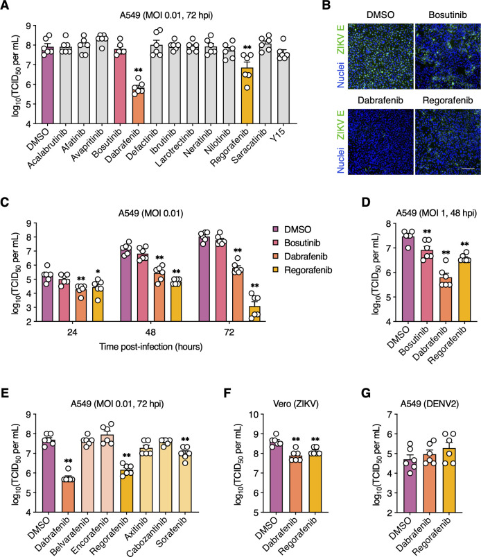
Dabrafenib and Regorafenib inhibit ZIKV but not DENV2 replication. (A and B) A549 cells were mock-infected or infected with ZIKV (MOI = 0.01 PFU/cell) and subsequently treated with the indicated SMKIs or DMSO (vehicle control). (A) Viral titers in the supernatant were determined at 72 hpi by endpoint dilution assay. (B) ZIKV infection was visualized at 72 hpi by immunofluorescence staining for ZIKV E protein (green). Nuclei were stained with Hoechst 33342 (blue). Scale bar, 200 µm. Representative images of two independent experiments are shown. (C) A549 cells were infected with ZIKV (MOI = 0.01 PFU/cell) and subsequently treated with DMSO, bosutinib (5 µM), Dabrafenib (10 µM), or Regorafenib (2.5 µM). Viral titers in the supernatant were determined at 2, 24, 48, and 72 hpi by endpoint dilution assay. (D) A549 cells were infected with ZIKV (MOI = 1 PFU/cell) and subsequently treated with DMSO, bosutinib (5 µM), Dabrafenib (10 µM), or Regorafenib (2.5 µM). Viral titers in the supernatant were determined at 48 hpi by endpoint dilution assay. (E) A549 cells were infected with ZIKV (MOI = 0.01 PFU/cell) and subsequently treated with the indicated SMKIs or DMSO. Viral titers in the supernatant were determined at 72 hpi by endpoint dilution assay. (F) Vero cells were infected with ZIKV (MOI = 0.01 PFU/cell) and subsequently treated with DMSO, Dabrafenib (2.5 µM), or Regorafenib (1 µM). Viral titers in the supernatant were determined at 48 hpi by endpoint dilution assay. (G) A549 cells were mock-infected or infected with DENV2 (MOI = 0.01 PFU/cell) and subsequently treated with DMSO, Dabrafenib (10 µM), or Regorafenib (2.5 µM). Viral titers in the supernatant were determined at 72 hpi by endpoint dilution assay. Data are expressed as mean ± standard error of the mean (SEM) from two independent experiments with three biological replicates per experiment. *, P < 0.05; **, P < 0.01 (Mann–Whitney U test vs “DMSO”).

