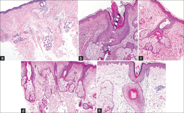
Figure 2.
Corresponding skin biopsies (All H and E, ×100, vertical sectioning) showing. (a) Case1 - Perifollicular inflammation and perifollicular fibrosis with follicular loss and relative expansion of sebaceous units, (b) Case 2 - Mild perivascular perifollicular lymphocytic inflammation and fibrosis with a reduction in the number of hair follicles giving a ‘pseudo sebaceous gland hyperplasia’appearance, (c) Case 3 - Subtle lichenoid inflammation is present at the level of the infundibulum with associated dilatation of the hair follicle, (d) Case 4 - Lichenoid inflammation affecting the vellus hair follicles at the level of the infundibulum, (e) Case 5 - Vellus hair follicular distortion with subtle inflammation and perifollicular fibrosis

