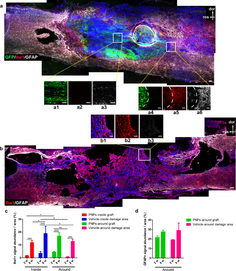
Fig. 8.
Gliosis at the lesion site two months after SCI with or without transplantation. (a, b) Staining results of microglia/macrophages (Iba1) and astrocytes (GFAP) in PNP group (a) and vehicle group (b) two months after SCI. (c, d) Quantification of Iba1 + signals (c) and GFAP + signals (d) at different areas of the lesion sites. n = 5; * P < 0.05; ** P < 0.01; *** P < 0.001. Iba1, red; GFAP, white; d, dorsal; r, rostral; bar, 100 μm in a and b; 50 μm in a1-a6 and b1-b3

