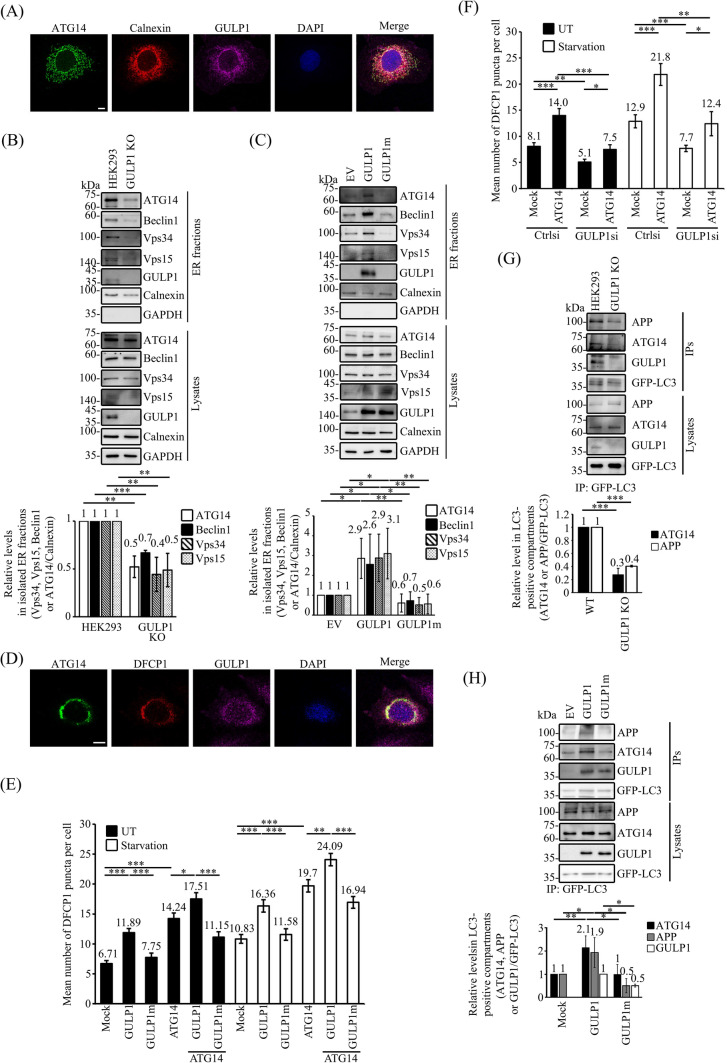
Fig. 4.
GULP1 facilitates the ER targeting of ATG14 and the recruitment of APP to autophagic vacuoles. A Immunostaining of COS7 cells for ATG14, calnexin and GULP1. ATG14, calnexin and GULP1 were stained by rATG14-2, 10427-2-AP and anti-GULP1 G-R3 respectively. An overlaid image is shown. Nuclei were stained with DAPI. Scale bar, 10 μm. B ER fractions were isolated from WT and GULP1-KO HEK293. Individual PI3KC3-C1 components in the isolated ER fractions were analyzed by immunoblotting by using anti-ATG14 PD026, anti-Beclin1 Bec-R3, anti-Vps34 F-11, anti-Vps15 JK-13 and anti-GULP1 G-R3, respectively. Subcellular compartment markers including calnexin and GAPDH were detected with anti-calnexin 10427-2-AP and anti-GAPDH AM4300 respectively. Data were obtained from three independent experiments. Bar chart shows the relative levels of PI3KC3-C1 components in GULP1-KO HEK293 cells compared to WT HEK293. **p < 0.01, ***p < 0.001. C ER fractions were isolated from EV, GULP1 and GULP1m stably transfected HEK293. Individual PI3KC3-C1 components in the isolated ER fractions were analyzed by immunoblotting by using anti-ATG14 PD026, anti-Beclin1 Bec-R3, anti-Vps34 F-11 and anti-Vps15 JK-13 respectively. Subcellular compartment markers including calnexin and GAPDH were detected with anti-calnexin 10427-2-AP and anti-GAPDH AM4300 respectively. Data were obtained from three independent experiments. Bar chart shows the levels of PI3KC3-C1 components relative to EV. *p < 0.05, **p < 0.01. D Immunostaining of CHO cells transfected with ATG14, mCherry-DFCP1 and GULP1. ATG14 and GULP1 were stained by rATG14-2 and an anti-GULP1 G-R3 respectively. An overlaid image is shown. Nuclei were stained with DAPI. Scale bar, 10 μm. E & F Quantification of mCherry-DFCP1-positive puncta plotted by different transfection and treatment as indicated. Data was obtained from at least 40 cells per transfection, and the experiment was repeated three times. Error bars are sem. *p < 0.05, **p < 0.01, ***p < 0.001. G WT and GULP1-KO HEK293 cells were transfected with GFP-LC3. Autophagic vacuoles were immunoprecipitated with anti-GFP JL-8 antibody. The protein content in total cell lysates and immunoisolated GFP-LC3 positive fractions was analyzed by anti-APP A5137, anti-ATG14 PD026 and anti-GULP1 G-R3. Bar chart shows the densitometric quantification of ATG14 and APP against GFP-LC3 in IPs. The experiment was repeated three times. ***p < 0.001. H Stable EV, GULP1 and GULP1m HEK293 cells were transiently transfected with GFP-LC3. Autophagic vacuoles were immunoprecipitated with anti-GFP JL-8 antibody. The protein content in total cell lysates and immunoisolated GFP-LC3 positive fractions was analyzed by anti-APP A5137, anti-ATG14 PD026 and anti-GULP1 G-R3. Bar chart shows the densitometric quantification of ATG14 and APP against GFP-LC3 in IPs. The experiment was repeated three times. *p < 0.05, **p < 0.01

