Fig. 5.
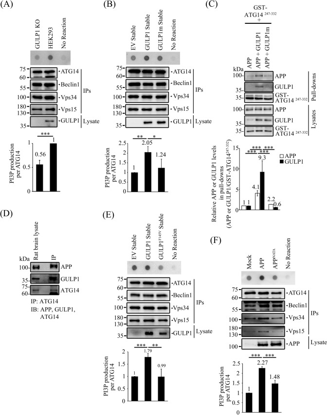
The GULP1-APP interaction enhances PI3KC3-C1 kinase activity. A WT and GULP1-KO HEK293 cells were transfected with ATG14, Beclin1, Vps34 and Vps15 and ATG14 was immunoprecipitated with anti-ATG14 anti-myc antibody 60003-2-IG. The immunoprecipitates were incubated with PI and ATP for 30 min. PI3P production was determined by dot blot and detected with GST-p40-phox. Bar chart shows the quantification of PI3P production normalized with immunoprecipitated ATG14. The experiment was repeated three times. ***p < 0.001. B Stable EV, GULP1 and GULP1m HEK293 cells were transfected with ATG14, Beclin1, Vps34 and Vps15 and ATG14 was immunoprecipitated with an anti-myc antibody 60003-2-IG. The immunoprecipitates were incubated with PI and ATP for 30 min. PI3P production was determined by dot blot and detected with GST-p40-phox. Bar chart shows the quantification of PI3P production normalized with immunoprecipitated ATG14. The experiment was repeated three times. ***p < 0.001. C CHO cells were transfected with GST-ATG14247−332 + APP, GST-ATG14247−332 + APP + GULP1 and GST-ATG14247−332 + APP + GULP1m. GST baits from cell lysates were captured by glutathione resins. Protein levels of APP, GULP1 and GST-ATG14247−332 were analysed with immunoblotting. Bar chart shows the densitometric quantification of co-precipitated APP and GULP1 relative to GST-ATG14247−332 baits. The experiment was repeated three times. ***p < 0.001. D ATG14 was immunoprecipitated with anti-ATG14 PD026 antibody from total rat brain lysate. APP, GULP1 and ATG14 in lysate and immunoprecipitates were analyzed by immunoblotting with anti-APP A5137, anti-GULP1 G-R3 and anti-ATG14 PD026. E Stable EV, GULP1 and GULP1F145V HEK293 cells were transfected with ATG14, Beclin1, Vps34 and Vps15 and ATG14 was immunoprecipitated with anti-myc antibody 60003-2-IG. The immunoprecipitates were incubated with PI and ATP for 30 min. PI3P production was determined by dot blot. Bar chart shows the quantification of PI3P production normalized with immunoprecipitated ATG14. The experiment was repeated three times. ***p < 0.001. F HEK293 cells were transfected with ATG14, Beclin1, Vps34 and Vps15 and ATG14 and mock, APP or APPNATA was immunoprecipitated with anti-myc antibody 60003-2-IG. The immunoprecipitates were incubated with PI and ATP for 30 min. PI3P production was determined by dot blot. Bar chart shows the quantification of PI3P production normalized with immunoprecipitated ATG14. The experiment was repeated three times. ***p < 0.001

