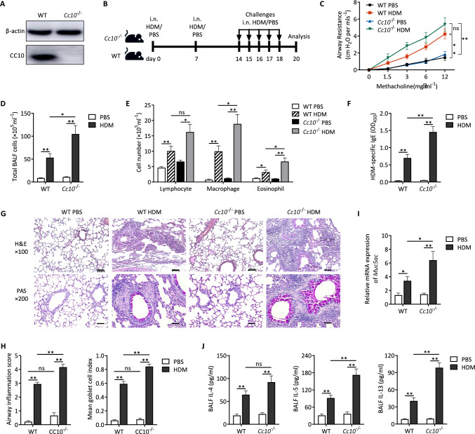
Fig. 2.
Enhanced allergic asthma in Cc10−/− mice post HDM exposure. A Western blot showing absence of CC10 in Cc10−/− lung tissue. B HDM sensitization/challenge protocol for wild type and Cc10−/− mice, with PBS controls. Analysis occurred 2 days post-final challenge. C Airway resistance to MCh increments; normalized to baseline (n = 6/group). D, E BALF cell counts (n = 6/group). F ELISA quantification of HDM-specific IgE (n = 6/group). G Lung histology stained with H&E or PAS; scale bar = 200 μm. H Quantitative assessment of inflammatory infiltration and goblet cell hyperplasia (n = 6/group). I Quantification analysis for Muc5ac mRNA expression in mice lung tissues (n = 4/group). J ELISA determination of IL-4, IL-5, and IL-13 in BALF (n = 6/group). Data are mean ± SEM. P values: one-way ANOVA with Games-Howell test (D–I), two-way ANOVA (C). *P < 0.05, **P < 0.01, ns not significant

